
Check out our latest products

![[5G & 2.4G] Indoor/Outdoor Security Camera for Home, Baby/Elder/Dog/Pet Camera with Phone App, Wi-Fi Camera w/Spotlight, Color Night Vision, 2-Way Audio, 24/7, SD/Cloud Storage, Work w/Alexa, 2Pack](https://m.media-amazon.com/images/I/71gzKbvCrrL._AC_SL1500_.jpg)


The hybrid microscope uses two technologies to capture molecules’ 3D position and orientation, helping scientists study biological structures more clearly.
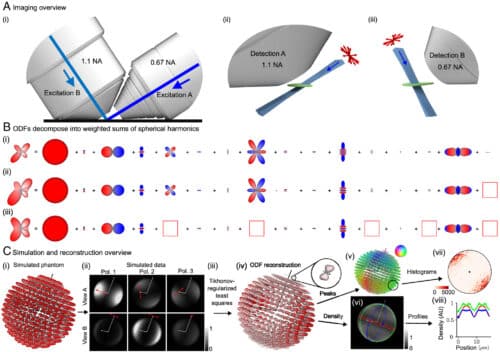
The idea that two are better than one applies not just to people but also to scientific instruments. By combining two technologies, researchers at the Marine Biological Laboratory (MBL) have developed a hybrid microscope that can, for the first time, capture both the full 3D orientation and position of molecules, such as labelled proteins inside cells.
The microscope integrates polarized fluorescence technology, which measures molecular orientation, with a dual-view light sheet microscope (diSPIM), which provides detailed imaging along a sample’s depth (axial) axis.

This combination has significant potential. Proteins, for instance, adjust their 3D orientation in response to their environment, enabling interactions with other molecules essential for their function.
Traditional microscopy, including polarized light, can effectively study the spindle when positioned perpendicular to the viewing direction. However, when the spindle is tilted, the readout becomes ambiguous. The new instrument addresses this limitation by correcting for tilt, allowing researchers to accurately capture spindle molecules’ 3D orientation and position, such as microtubules.
Imaging molecules within the spindle of a dividing cell—a challenge at MBL and beyond—is another key application. The team aims to improve the system’s speed to track changes in the position and orientation of structures in live samples over time. They also hope that future fluorescent probes will expand their use to a wider range of biological structures.
A Confluence of Vision
The diSPIM microscope uses two imaging paths that intersect at a right angle, enabling researchers to illuminate and capture the sample from both perspectives. This dual view improves depth resolution and provides greater polarisation control than other microscopes.
The researchers believe that this setup could also overcome a key limitation of polarized light microscopy: the challenge of efficiently illuminating a sample with polarized light along the direction of light propagation.
Reference: Volumetric imaging of the 3D orientation of cellular structures with a polarized fluorescence light-sheet microscope. Proceedings of the National Academy of Sciences, 2025; 122 (8) DOI: 10.1073/pnas.2406679122

![[3 Pack] Sport Bands Compatible with Fitbit Charge 5 Bands Women Men, Adjustable Soft Silicone Charge 5 Wristband Strap for Fitbit Charge 5, Large](https://m.media-amazon.com/images/I/61Tqj4Sz2rL._AC_SL1500_.jpg)