Check out our latest products
Researchers have developed a technique for advanced microscopy using an ultra-thin optical fibre, opening new frontiers in medical imaging.

Scientists at the University of Adelaide, Australia in collaboration with international partners, have pioneered a technique for advanced microscopy through an optical fibre thinner than a human hair. The breakthrough could transform medical imaging by allowing access to previously unreachable areas of the human body while minimising tissue damage.
Dr Ralf Mouthaan, the centre of light for life, University of Adelaide, explained that conventional methods faced limitations due to distortions occurring as light propagated through hair-thin optical fibres. These distortions resulted in random granular patterns, rendering them unsuitable for complex microscopy. “Performing advanced microscopy in a hair-thin fibre will reveal a wealth of additional information,” Dr Mouthaan noted.

The innovation holds particular promise for medical professionals, researchers in nanotechnology, and biomedical engineers who require highly detailed imaging of delicate or inaccessible structures. By addressing existing limitations, this technique opens avenues for diagnosing diseases, studying biological processes, and exploring nanoscale materials.
Using innovative approaches, the team pre-shaped light to generate precise optical patterns, overcoming the challenges of distortion. Techniques like light sheet microscopy and stimulated emission-depletion (STED) microscopy stand to benefit immensely. STED microscopy, for instance, allows imaging of nanostructures with diameters measured in billionths of a metre.
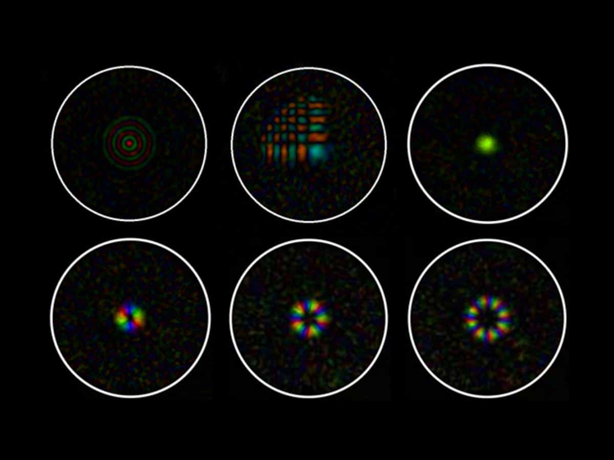
The researchers demonstrated the ability to project intricate patterns such as Bessel beams, Airy beams, and Laguerre-Gaussian beams through a fibre core of just 50µm. “There is almost no limit to what can be projected through the fibre. For example, a letter such as the Greek alpha can also be formed,” Dr Mouthaan added.
The project involved collaboration with experts from the University of Nottingham and the University of Cambridge, alongside Kishan Dholakia, professor and director of the centre of light for life. The research provides unprecedented control over beam properties like amplitude, phase, and polarisation.
Looking ahead, the team plans to create “endomicroscopes” to provide proof of concept, while partners at Nottingham work on clinically applicable endoscopes. By miniaturising advanced microscopy, this innovation heralds a new era in medical diagnostics and research.


![[5G & 2.4G] Indoor/Outdoor Security Camera for Home, Baby/Elder/Dog/Pet Camera with Phone App, Wi-Fi Camera w/Spotlight, Color Night Vision, 2-Way Audio, 24/7, SD/Cloud Storage, Work w/Alexa, 2Pack](https://m.media-amazon.com/images/I/71gzKbvCrrL._AC_SL1500_.jpg)


![[3 Pack] Sport Bands Compatible with Fitbit Charge 5 Bands Women Men, Adjustable Soft Silicone Charge 5 Wristband Strap for Fitbit Charge 5, Large](https://m.media-amazon.com/images/I/61Tqj4Sz2rL._AC_SL1500_.jpg)


